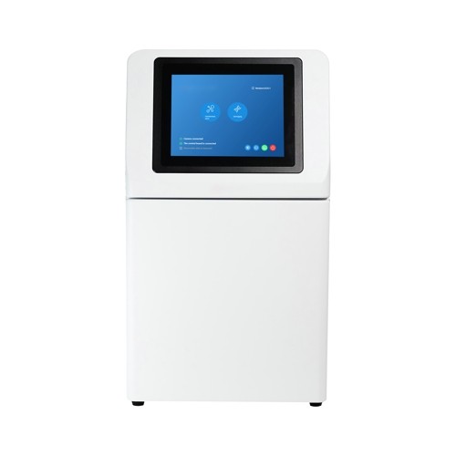

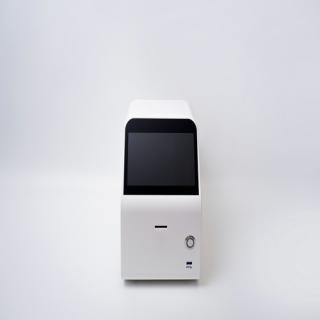
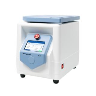
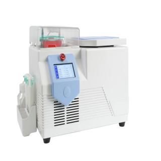
Multi-functional Imaging System
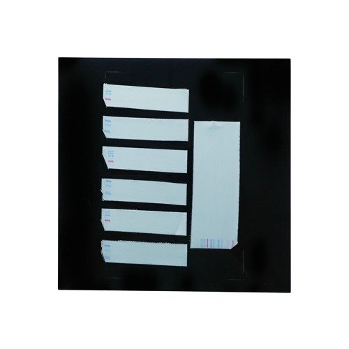
Integrated with a chemiluminescence system and a gel imaging system, it can be used for experiments such as Western Blotting membrane detection, nucleic acid gel imaging/gel cutting, and protein gel imaging.
Product Information
The KB-SCG-W5000 is a comprehensive device that integrates chemiluminescence technology and gel imaging. it is
equipped with a high-sensitivity cooled camera with 9 million pixels, enabling rapid, accurate, and high
throughput detection and imaging of samples. it is widely used in the fields of life sciences, medicine, and
environmental protection.
Product Features
Chemiluminescence Imaging System :
1. Weak band signal, one-time imaging
2. Both strong and weak band signals can be achieved, and the desired results can be obtained
3. Source file can be saved, and the image can be adjusted at any time
4. High Efficient
Gel Imaging System:
Three modes and multiple parameters can be flexibly adjusted, resulting in clear and bright imaging
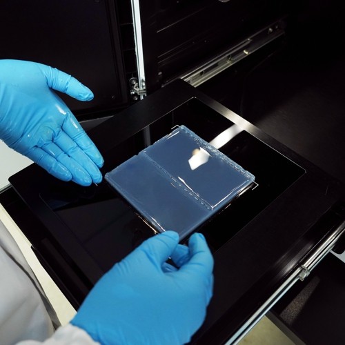
UV laser protective board, safe and convenient for gel cutting
TechnicalSpecifications
| Product Name | Multi-functional Imaging System | |
| Cat.No. | KB-SCG-W5000 | |
| Dimensions | 400×371×700 mm | |
| Camera | PixelResolution | 9 million |
| Resolution | 3000×3000 | |
| Pixel size | 3.76×3.76μm | |
| Target size | 1“(11.28×11.28 mm) | |
| Full Well Capacity | 16.5ke-(HCG),50.5ke-(LCG) | |
| Sensitivity | 877mv@1/30s | |
| ReadoutNoise | 1.24e-(HCG),3.22e-(LCG) | |
| Dark Current | 0.0003e-/s/pixel@-15℃ | |
| Signal-to-Noise Ratio | 42.2dB(HCG),47dB(LCG) | |
| ExposureTime | 0.1ms~1h | |
| Binning Mode | 1×1,2×2,3×3 | |
| Grayscale | 16-bit(65536 levels) | |
| Cooling | Relative to Ambient Temperature -40°c | |
| Camera Type | Black and White Camera | |
| Lens | Aperture | F0.95-F16 |
| Focal Length | 17mm | |
| Type | Motorized zoom lens | |
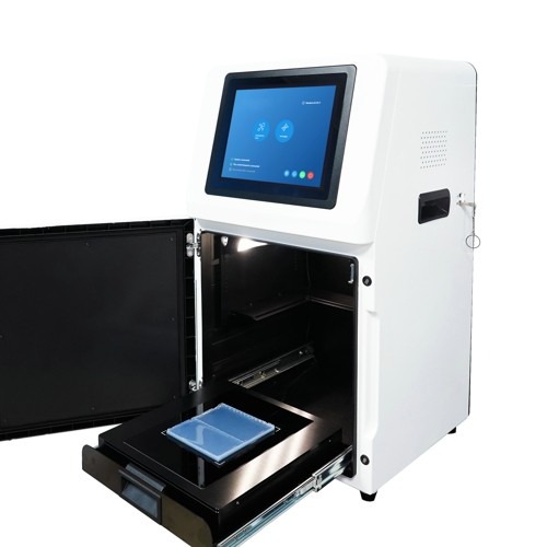
Light Source | Bright Field Light source | Downward-facing LED white light source, symmetrically distributed on both sides |
| Ultraviolet light source | 310nm LED array, providing uniform transmissive illumination. | |
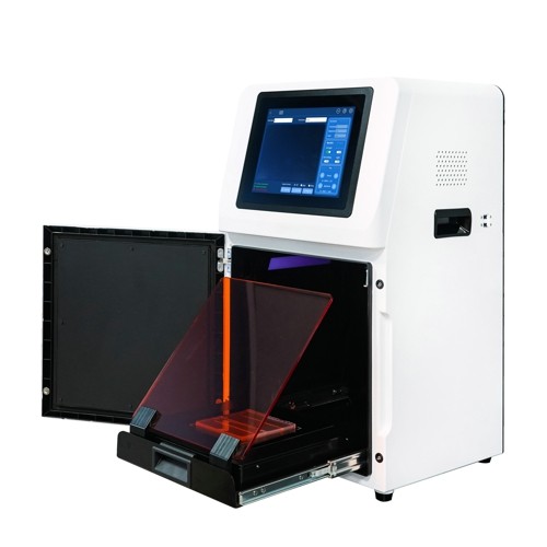
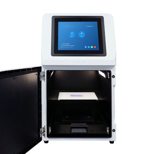
| Dark Box | Light isolation | Fully light-sealed, isolates environmental light. |
| Door Control | The door control sensor can control the on/off of the bright field light source. | |
| Rotating disc | Switch the filter according to the current mode to match the applications of chemiluminescence and gelimaging. | |
| Field of View | Effective field of view for membraneimagingis136mmxl36mm (expandable to200mmx200mm if necessary) Effective field of view for proteingelimagingis136mmx 136mm (expandableto200mmx 200mmifnecessary) The effective field of view for nucleic acid gel imaging is 200mmx200mm | |
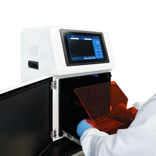
| Gel Cutting | After opening the door, the UV light source can be extracted and used with aUV protective board for cutting adhesive | |
Software Functions | Exposure Modes | High Quality: Image quality is the highest |
| Auto Exposure | Intelligent exposure technology quickly determines the optimal exposure time. With the combination of time imaging and time accumulation functions, users can achieve the best image results with just one operation. | |
| Real-time imaging | Real-time presentation of the changes in sample signals during the exposure process. allowing for the observation of every detail of the capture. Overexposed areas will be indicated for samples with overexposure. | |
| Time imaging | After exposure is complete, each frame image within the exposure time can be generated Through precise retrospective adjustments, users can choose any frame image within that exposure time as the final output. | |
| Time Accumulation | For samples with insufficient exposure, users can choose to continue exposure after the initial exposure is completed, enabling the sample to receive additional exposure on top of the already exposed time. | |
| IndustrialComputer | 10.4 inches,1024x768,Windows operating system | |
| ExternalInterfaces | USB 3.0×2 | |
| Operating Voltage | 100V-240V | |
| ProductPower | 100W | |
| Product Net Weight | 30Kg | |
| Cat. No. | SCG-W5000 | SCG-W3000 | SCG-W1000 |
| Dimension | 400×371×700 mm | 400×371×700 mm | 400×371×700 mm |
| Camera | Depth-cooled high sensitivity camera | Depth-cooled high sensitivity camera | High-sensitivity camera |
| Resolution | 2992*3000,9 megapixels | 2992*3000, 9 megapixels | 3072*2048,4.2 megapixels |
| Pixel | 3.76×3.76 μm | 3.76×3.76 μm | 2.4×2.4 μm |
| Shooting Area | Effective field of view for blotting film/protein gel: 136×136 mm (can be expanded to 200×200 mm if required); Effective field of view for nucleic acid gel: 200×200 mm. | Blotting Film 136×136 mm (expandable to 200×200 mm if required) | Nucleic Acid Gel / Protein Gel 140×140 mm |
| Cooling Temperature | Relative ambient temperature -40°C | Relative ambient temperature -40°C | - |
| Light Source | Bright-field Light Source: Downward-facing LED white light source, symmetrically distributed on both sides. UV Light Source: 310 nm LED array for uniform transmission illumination. | Downward-facing LED white light, symmetrically distributed on both sides | Bright-field Light Source: Downward-facing LED white light source, symmetrically distributed on both sides. UV Light Source: 310 nm LED array for uniform transmission illumination. |
Inquiry
Product
CATALOGUE
 sales
sales